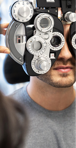
You may have heard your optometrist recommend OCT imaging during your eye exam, but what is it really? OCT is a powerful tool your optometrist uses to monitor your eye health and diagnose eye disease.
3D Digital Eye Exams
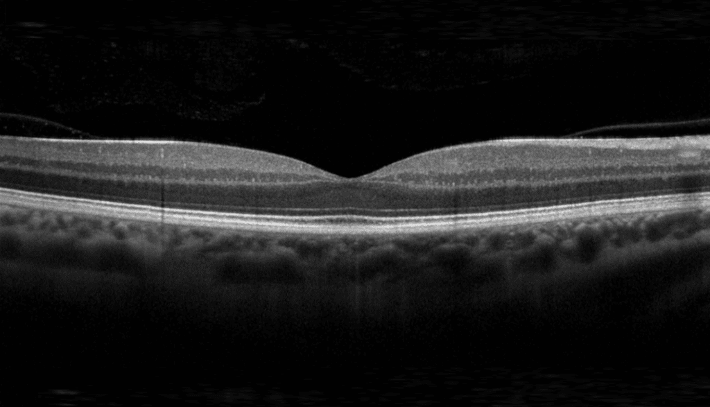
Optical coherence tomography, or OCT for short, is sometimes referred to as a “3D” or “digital” eye exam. That’s because OCT involves taking 3D, digital images of your eye. Specifically, OCT takes a cross-sectional image of your retina, the light-sensitive part of your eye.
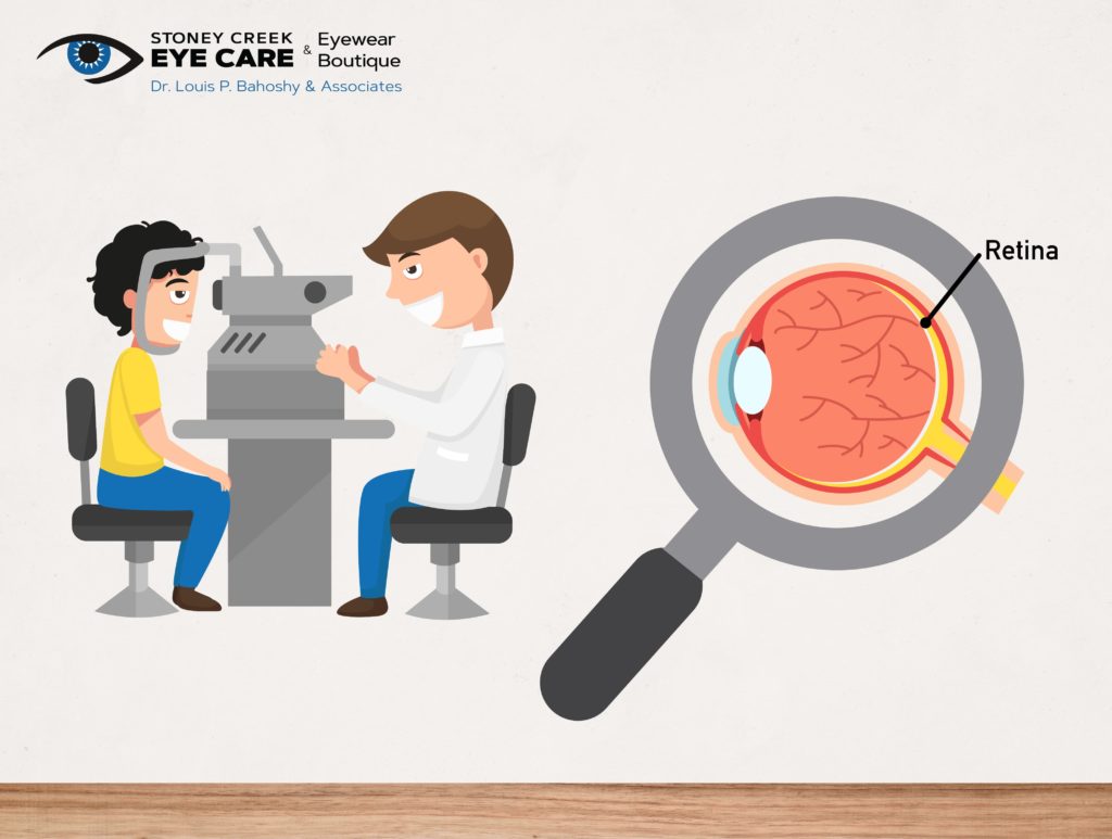
How Does OCT Work?
OCT works similarly to an ultrasound. It provides real-time images of your eye’s internal structures like an ultrasound, but OCT uses light waves instead of sound waves. These light waves illuminate and scan your retina, giving your optometrist a detailed view. So detailed, that your optometrist can evaluate your retina at a near-cellular resolution.
What to Expect During an OCT Exam
From start to finish, an OCT exam takes about 10 minutes. The exam is non-invasive and painless. You simply place your chin into a chin rest and keep your eye open as you look at a target (often a blinking dot or a small picture). Then, without touching it, the OCT machine scans your eye.
Dilation
Depending on your risk level or history of eye disease, your optometrist might recommend that your eyes are dilated during the OCT exam. Dilating your eyes is a simple procedure and gives your optometrist an even larger view of your retina.
If you’re undergoing dilation, your optometrist will put eye drops into your eyes that cause your pupils to expand, or dilate. Most patients find dilation painless, although some light sensitivity is to be expected. You’ll still be able to drive yourself home from the exam, but make sure you bring a pair of sunglasses to help with the light sensitivity.
Benefits of OCT Testing
Non-Invasive
OCT testing gives your optometrist an in-depth look into your eyes without feeling in-depth. The process is quick and painless, with nothing touching your actual eye. It’s no more hassle than getting a school photo done, except in the case of OCT, the photo is of your retina.
Early Disease Diagnosis
OCT is a great tool in diagnosing eye disease. In some cases, OCT can help your optometrist diagnose eye disease earlier, which allows for more effective treatment. Because OCT images give your optometrist such a detailed view of your retina, they can detect small indications of eye disease.
In addition to early disease diagnosis, OCT can also help with:
- Monitoring the progression of eye disease.
- Diagnosing eye disease in children, which can otherwise be difficult.
- Diagnosing other diseases like high blood pressure, multiple sclerosis, and vascular diseases.
Do I Need an OCT Exam?
Your optometrist will likely recommend an OCT exam if you have an increased risk of eye disease. You probably have an increased risk if you are over age 50 or have:
- A personal history of certain eye conditions
- A family history of eye disease
- Diabetes
- High blood pressure (hypertension)
Even if you don’t have a high risk of eye disease, OCT exams are still a great way to help protect your eye health. By having OCT exams regularly, it creates a baseline that your optometrist can reference. Using this baseline, your optometrist can detect the smallest changes to your eyes over time.
What Diseases Can OCT Help Diagnose?
Age-Related Macular Degeneration
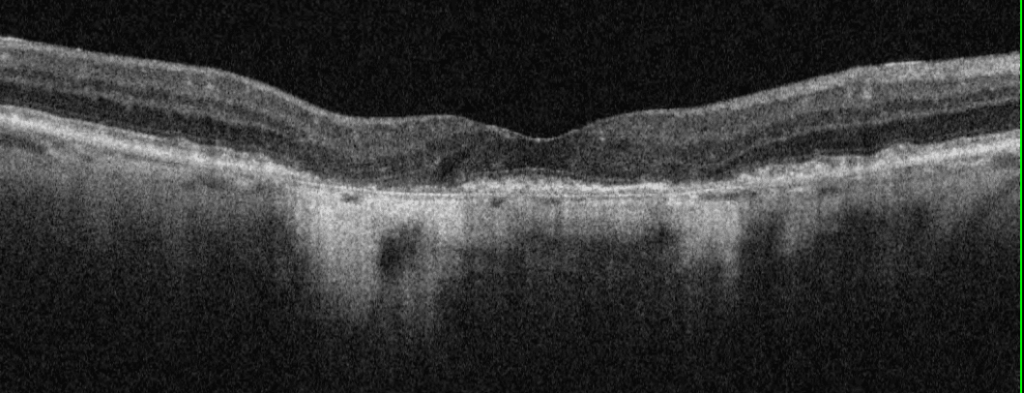
Age-related macular degeneration (AMD) is one disease OCT can help diagnose, often earlier than other methods. AMD affects the macula, which is the central part of the retina.
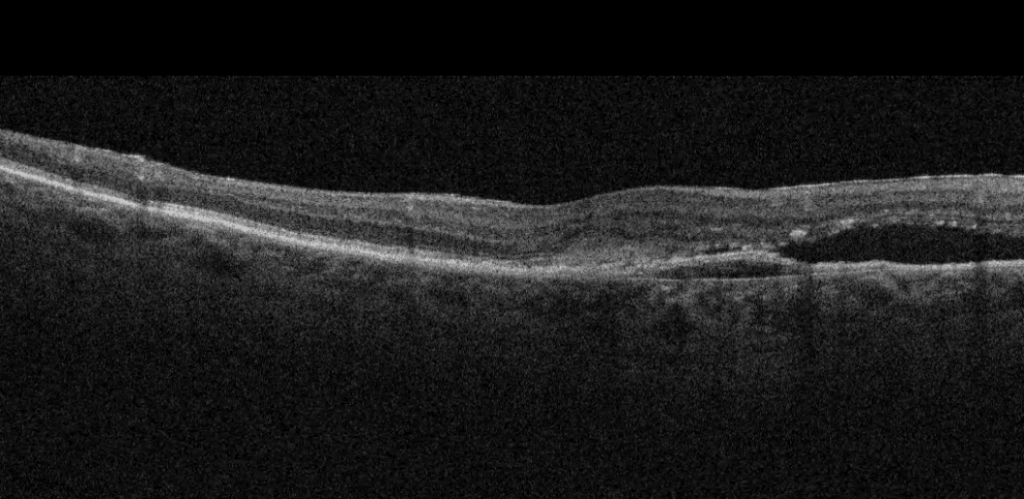
OCT provides detailed images of the retina and macula, showing irregularities caused by AMD. With OCT imaging, your optometrist can detect signs of AMD like drusen (tiny clumps of protein) caused by dry AMD and abnormal blood vessels and bleeding caused by wet AMD.
Glaucoma
In addition to producing detailed images of the retina, OCT can also show the optic nerve, which is the part of the eye affected by glaucoma. OCT can help with diagnosing glaucoma by showing damage and changes to the optic nerve.
In particular, OCT can help diagnose forms of glaucoma that don’t cause increased intraocular pressure and therefore, can’t be diagnosed with standard glaucoma tests.
Diabetic Retinopathy
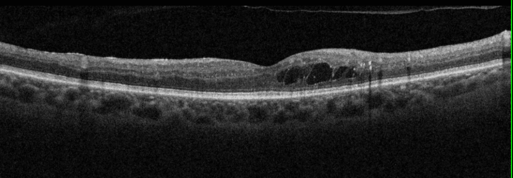
Diabetic retinopathy causes damage to the retina, specifically to the blood vessels. OCT imaging shows these damaged or abnormal blood vessels, allowing for diagnosis.
Other Problems in the Retina & Macula
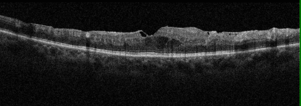
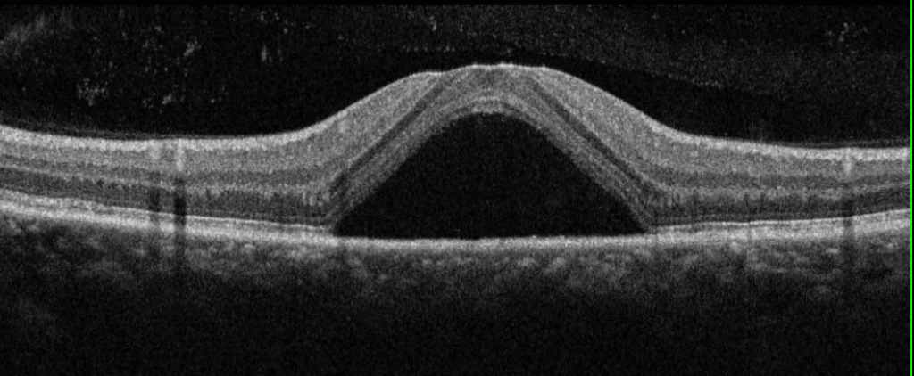
Because OCT gives such detailed images of the retina and macula, it can help diagnose a variety of other conditions, including:
- Retinal tear
- Retinal detachment
- Central serous retinopathy
- Retinal/macular cysts
- Macular hole
- Macular pucker
- Macular edema
- Other conditions that change how light passes through the eye (like cataracts and vitreous bleeding).
OCT provides your optometrist with a wealth of information about your eye health. Whether you’re at risk for eye disease or simply having a routine eye exam, OCT images are a valuable tool that provide valuable insight.


